Product : Book
Language : English
Cover : Paperback (Fig 1)
Quality of paper and images : The highest (Fig 2)
No. of pages : 518 (Fig 3)
No. of videos : 1,100 (Video 1)
Publisher : OMF Publishing LLC
Published : 2017
ISBN : 978-966-97590-3-0
Item weight : 1,100 grams (i.e., 38.8 ounces)
Dimensions : 15.1 x 2.5 x 22.5 cm (i.e., 5.94 x 0.98 x 8.85 inches)
Book`s Instagram : @essential.radiology
About the book on the ResearchGate website : Link
YouTube Channel of the Book : Link


FIGURE 1. Cover of the book titled "Essential Radiology for Medical Students, Interns and Residents": Paperback.

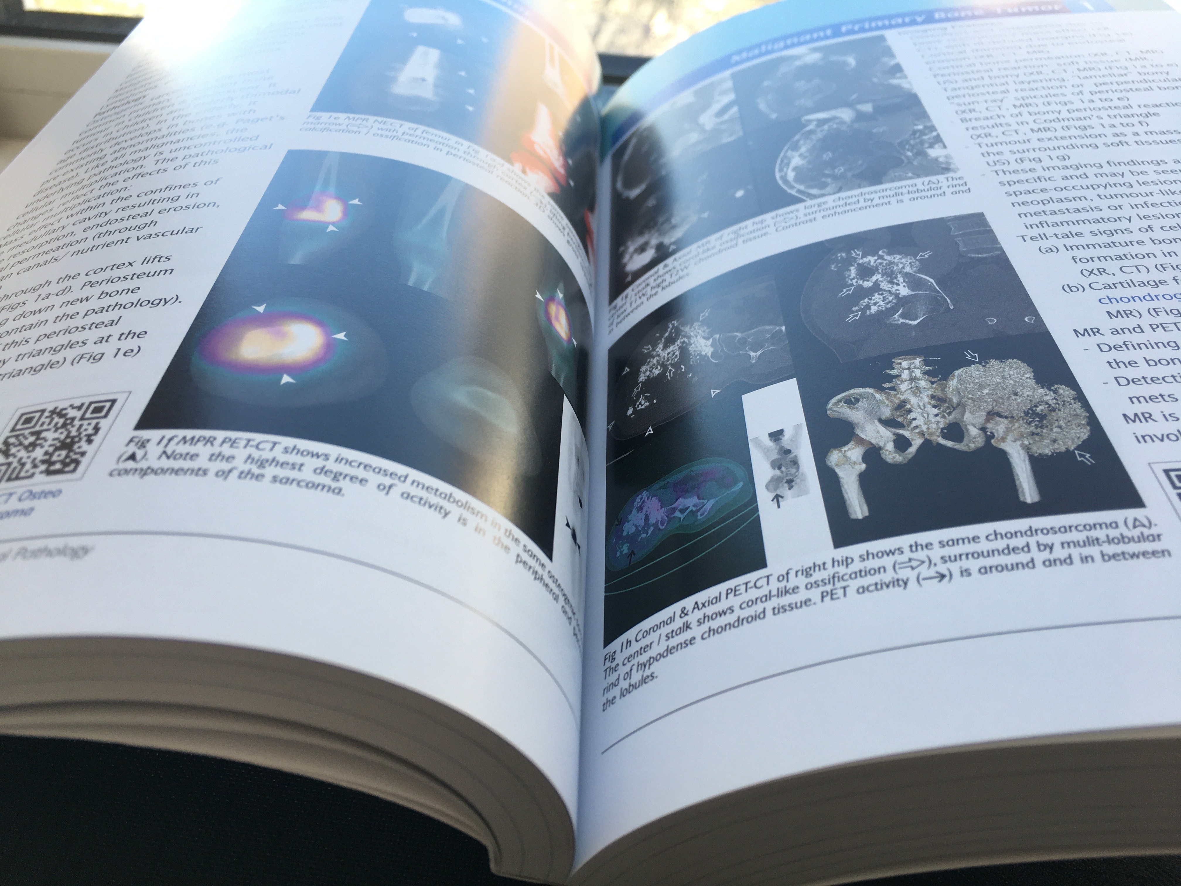
FIGURE 2. The book, titled "Essential Radiology for Medical Students, Interns and Residents" has excellent printing, high quality radiological images and educational material.
PURCHASE AND DELIVERY
Purchase of one book copy with delivery outside Ukraine : 145 euro (i.e., 154 USD or 122 GBP), the price includes delivery to any country (cost as of April 24, 2024).
The average time of delivery of a book from Ukraine to the farthest countries from it : 14 days. Delivery time confirmation (download PDF).
To make an order please contact us via the E-mail : office@omfpublishing.com.

Purchase of three (3) book copies with delivery outside Ukraine : 115 euro (i.e., 123 USD or 97 GBP) for each copy of the book + 42 euro (delivery). That is, the total cost of three (3) copies of the book with delivery will be 387 euros (i.e., 414 USD or 326 GBP). Cost as of April 24, 2024.
Purchase of five (5) book copies with delivery outside Ukraine : 115 euro (i.e., 123 USD or 97 GBP) for each copy of the book + 42 euro (delivery). That is, the total cost of five (5) copies of the book with delivery will be 617 euros (i.e., 659 USD or 519 GBP). Cost as of April 24, 2024.
We are an Ukraine-based publishing house and we are happy to contribute to the growth of your library.


FIGURE 3. The thickness of the book titled "Essential Radiology for Medical Students, Interns and Residents" is 518 pages.
DESCRIPTION
This is a comprehensive system-based review in clinical radiology, covering major diseases encountered in everyday clinical practice. A case-orientated approach is used, with high quality images from the latest available imaging modalities including MRI of the breast and cardiovascular system, CT colonography, amongst others. Each chapter is dedicated to a specific functional system and begins by outlining different clinical cases, each with a concise clinical history and initial imaging, followed by explanation of the pertinent imaging findings. The second part of each case contains a discussion on the list of important differential diagnoses to consider and the role of different imaging modalities in the presenting problem. The latter shows how information derived from different imaging modalities (including xrays, fluoroscopy, CT, MRI, nuclear medicine / PET-CT, angiograms, etc.) can help refine and prioritize imaging differentials. For each clinical case, the novel incorporation of cine images (accessible via QR codes) provides a new type of learning experience – one that simulates the reality of how medical imaging are int erpreted using serial images with spatial reformatting rather than static planar images.
TABLE OF CONTENTS
Front cover (download PDF)
Contents (download PDF)
Affiliation / Acknowlegment (download PDF)
Preface (download PDF)
Dedication (download PDF)
-
Section 1 : Head & Neck. This Section includes 11 Chapters. The Section 1 is open access, publicly available and free to download. The PDF file has 50 pages and occupies 28 MB. Download PDF.
-
Section 2 : Genitourinary System.
-
Section 3 : Hepatobiliary System.
-
Section 4 : Tumor & Tumor-like Musculoskeletal Pathology.
-
Section 5 : Joint Pathology & Trauma.
-
Section 6 : Central Nervous System.
-
Section 7 : Chest.
-
Section 8 : Cardiovascular System.
-
Section 9 : Breast.
-
Section 10 : Gastrointestinal System.
VIDEO 1. A sample of one of the integrated videos due to QR codes. Multiplanar reformation (MPR) (reconstruction) and surface rendered non-enhanced computed tomography (NECT) shows chronic septic arthritis, with cartilage and bone destruction/ resorption, widening of intercondylar groove, lateral subluxation on patella at patellofemoral joint (PFJ). Section 5 : Joint Pathology and Trauma. Chapter 3 : Acute Septic Arthritis.
This book is available in the libraries of such universities as:
-
Bogomolets National Medical University, Ukraine.
-
Kyiv Medical University, Private Higher Educational Establishment, Ukraine (Fig 2).
-
The Chinese University of Hong Kong, Hong Hong.
This book is available in state institutions:
-
Ivan Fedorov Book Chamber of Ukraine, State Scientific Institution, Ukraine.
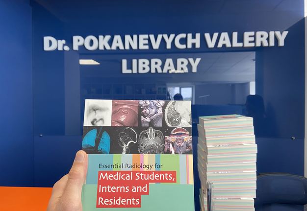

FIGURE 4. The book, titled "Essential Radiology for Medical Students, Interns and Residents" and the Dr. Pokanevych Valeryi Library, Kyiv Medical University, Private Higher Educational Establishment, Kyiv, Ukraine.
REVIEWS
“After a hard wok, it is finally here: radiologists deeply specialized in the diagnostics of head and neck disorders give the readers a possibility to touch the cutting-edge practical book Essential Radiology for Medical Students, Interns and Residents. The textbook is edited by Professor Ahuja AT, the most experienced world radiologist from Hong Kong (SAR).
Content consists of ten Sections. 1st of which, Head & Neck, is critically important to the specialists related with that area of human body. Uniqueness of that work – it consists of 1100 cine loops (CT, MRI scans, US images, etc.) connecting via QR codes. And to Head & Neck Section belongs 111 of them covering the whole range of pathologic conditions.
In summary, I would not hesitate to recommend this book to anyone interested in making diagnosis as precise as possible.” (Journal of Diagnostics and Treatment of Oral and Maxillofacial Pathology, 28 December 2017). PDF of the book review. Review link: https://doi.org/10.23999/j.dtomp.2017.3-4.5
ABOUT THE BOOK EDITOR
Dr. Anil T. Ahuja (Fig 5) MBBS (Mumbai), MD (Mumbai), FRCR (UK), FHKCR, FHKAM (Radiology)
Professor of Diagnostic Radiology & Organ Imaging
Department of Imaging & Interventional Radiology
The Chinese University of Hong Kong
Hong Kong, SAR.
Professor Anil T. Ahuja obtained his Radiology training in India (Seth G.S. Medical College, K.E.M Hospital & B.Y.L. Nair Hospital, Mumbai) and England and began his academic career at the Department of Imaging & Interventional Radiology of the Faculty of Medicine, Chinese University of Hong Kong in 1988.
Professor Ahuja has a keen interest in the imaging of Head and Neck diseases. His research is focused on Neck Masses, Lymph Nodes, Thyroid, Parathyroid, Salivary Gland and Head & Neck Tumours.
Professor Ahuja has published extensively in peer reviewed medical journals, lectured at international conferences, held hands-on workshops and is Assistant Editor for Clinical Radiology. He has edited more than ten books in Radiology and authored numerous book chapters.
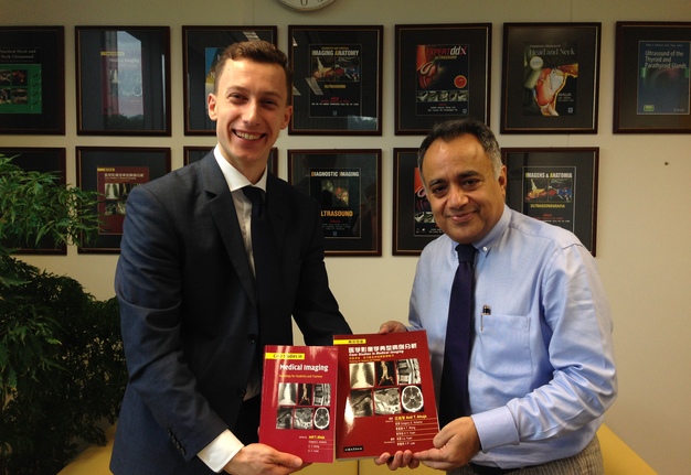
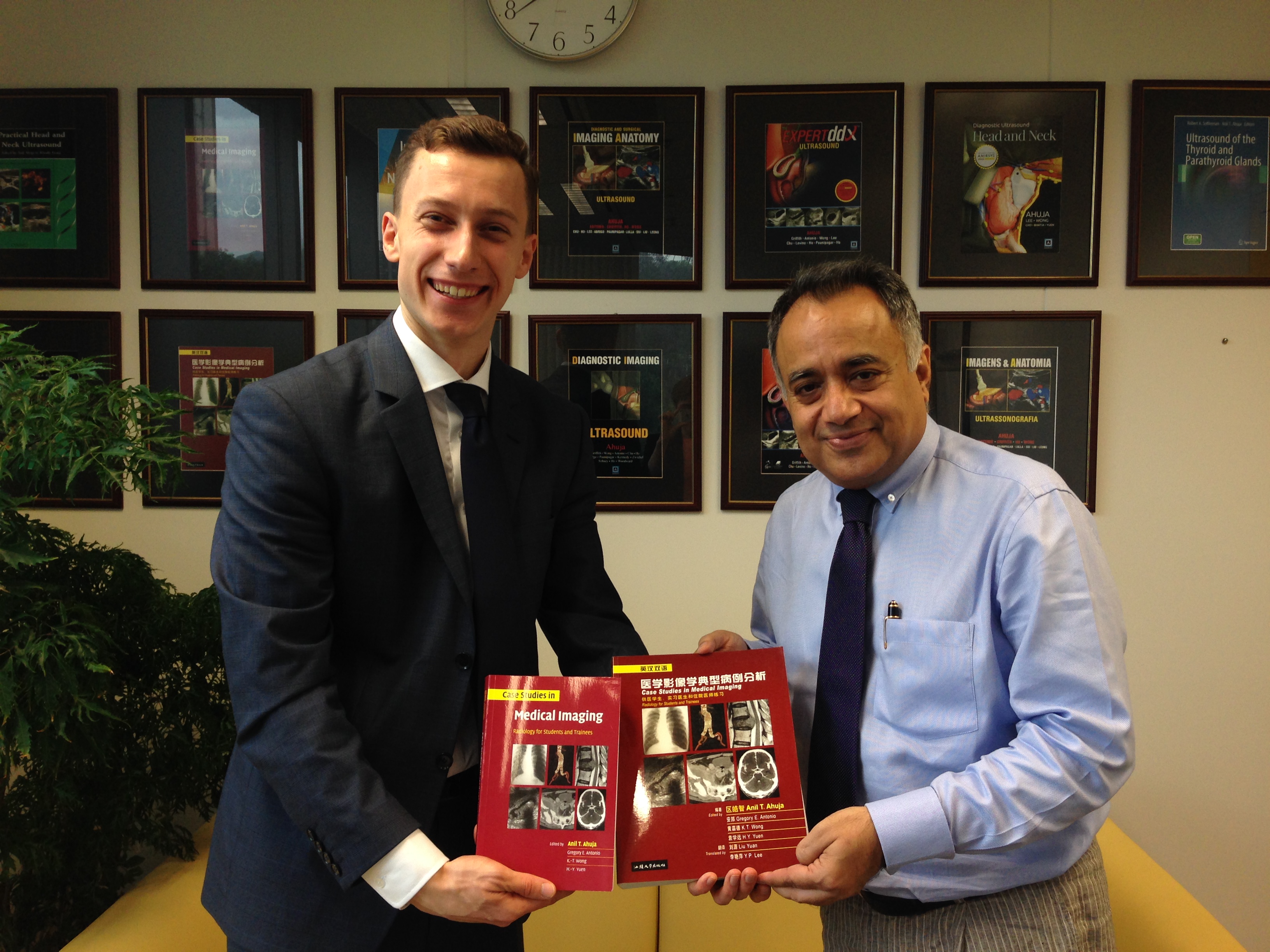
FIGURE 5. The representative of OMF Publishing LLC, Eugene Fesenko (left), and Professor Anil T. Ahuja (right) hold in their hands the previous editions of the Professor's team. Location: The Chinese University of Hong Kong, Hong Kong. Date: March, 2017.
BUYING A BOOK WITHIN UKRAINE. КУПІВЛЯ КНИГИ В МЕЖАХ УКРАЇНИ
Book purchase with delivery within Ukraine : The cost of the book with delivery within Ukraine is 110 euro (i.e., 4,630 UAH as of April 24, 2024).
Купівля книги з доставкою по Україні : Вартість книги з доставкою по Україні – 110 євро (тобто 4630 грн станом на 24.04.2024).
Щоб зробити замовлення, зв'яжіться з нами через електронну пошту office@omfpublishing.com.
КНИГА АНГЛІЙСЬКОЮ МОВОЮ. ОПИС КНИГИ
Це комплексний системний огляд клінічної радіології (променевої діагностики), що охоплює основні захворювання, які зустрічаються в повсякденній клінічній практиці. Використовується підхід, орієнтований на випадки, з високоякісними зображеннями з найновіших доступних методів візуалізації, включаючи МРТ молочної залози та серцево-судинної системи, КТ-колонографію тощо. Кожна глава присвячена певній функціональній системі та починається з опису різних клінічних випадків, кожен із короткою клінічною історією та початковою візуалізацією, після чого йдуть пояснення відповідних результатів візуалізації. Друга частина кожного випадку містить обговорення списку важливих диференційних діагнозів, які слід враховувати, і ролі різних методів візуалізації в даній проблемі. Остання показує, як інформація, отримана з різних методів візуалізації (включно з рентгенографією, флюороскопією, КТ, МРТ, ядерною медициною/ПЕТ-КТ, ангіограмами тощо), може допомогти уточнити та визначити пріоритетність відмінностей зображень. Для кожного клінічного випадку нове включення кінозображень (доступних за допомогою QR-кодів) забезпечує новий тип навчання – такий, який імітує реальність інтерпретації медичних зображень за допомогою серійних зображень із просторовим переформатуванням, а не статичних плоских зображень.